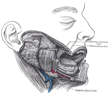
| is irregular in form and about the size of a walnut. A considerable part of it is situated in the submaxillary triangle, reaching forward to the anterior belly of the Digastricus and backward to the stylomandibular ligament, which intervenes between it and the parotid gland. Above, it extends under cover of the body of the mandible; below, it usually overlaps the intermediate tendon of the Digastricus and the insertion of the Stylohyoideus, while from its deep surface a tongue-like deep process extends forward above the Mylohyoideus muscle. | 128 |
| Its superficial surface consists of an upper and a lower part. The upper part is directed outward, and lies partly against the submaxillary depression on the inner surface of the body of the mandible, and partly on the Pterygoideus internus. The lower part is directed downward and outward, and is covered by the skin, superficial fascia, Platysma, and deep cervical fascia; it is crossed by the anterior facial vein and by filaments of the facial nerve; in contact with it, near the mandible, are the submaxillary lymph glands. | 129 |
 |
Saturday, April 17, 2010
Submaxillary Gland (glandula submaxillaris
Subscribe to:
Post Comments (Atom)


0 comments:
Post a Comment