| e tongue is partly invested by mucous membrane and a submucous fibrous layer. | 102 |
| The mucous membrane (tunica mucosa linguæ) differs in different parts. That covering the under surface of the organ is thin, smooth, and identical in structure with that lining the rest of the oral cavity. The mucous membrane of the dorsum of the tongue behind the foramen cecum and sulcus terminalis is thick and freely movable over the subjacent parts. It contains a large number of lymphoid follicles, which together constitute what is sometimes termed the lingual tonsil. Each follicle forms a rounded eminence, the center of which is perforated by a minute orifice leading into a funnel-shaped cavity or recess; around this recess are grouped numerous oval or rounded nodules of lymphoid tissue, each enveloped by a capsule derived from the submucosa, while opening into the bottom of the recesses are also seen the ducts of mucous glands. The mucous membrne on the anterior part of the dorsum of the tongue is thin, intimately adherent to the muscular tissue, and presents numerous minute surface eminences, the papillæ of the tongue. It consists of a layer of connective tissue, the corium or mucosa, covered with epithelium. | 103 |
| The epithelium is of the stratified squamous variety, similar to but much thinner than that of the skin: and each papilla has a separate investment from root to summit. The deepest cells may sometimes be detached as a separate layer, corresponding to the rete mucosum, but they never contain coloring matter. | 104 |
| The corium consists of a dense felt-work of fibrous connective tissue, with numerous elastic fibers, firmly connected with the fibrous tissue forming the septa between the muscular bundles of the tongue. It contains the ramifications of the numerous vessels and nerves from which the papillæ are supplied, large plexuses of lymphatic vessels, and the glands of the tongue. | 105 |
| Structure of the Papillæ.—The papillæ apparently resemble in structure those of the cutis, consisting of cone-shaped projections of connective tissue, covered with a thick layer of stratified squamous epithelium, and containing one or more capillary loops among which nerves are distributed in great abundance. If the epithelium be removed, it will be found that they are not simple elevations like the papillæ of the skin, for the surface of each is studded with minute conical processes which form secondary papillæ. In the papillæ vallatæ, the nerves are numerous and of large size; in the papillæ fungiformes they are also numerous, and end in a plexiform net-work, from which brush-like branches proceed; in the papillæ filiformes, their mode of termination is uncertain. | 106 |
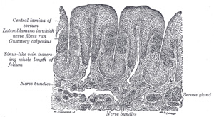
| Glands of the Tongue.—The tongue is provided with mucous and serous glands. | 107 |
| The mucous glands are similar in structure to the labial and buccal glands. They are found especially at the back part behind the vallate papillæ, but are also present at the apex and marginal parts. In this connection the anterior lingual glands (Blandin or Nuhn) require special notice. They are situated on the under surface of the apex of the tongue (Fig. 1013), one on either side of the frenulum, where they are covered by a fasciculus of muscular fibers derived from the Styloglossus and Longitudinalis inferior. They are from 12 to 25 mm. long, and about 8 mm. broad, and each opens by three or four ducts on the under surface of the apex. | 108 |
| The serous glands occur only at the back of the tongue in the neighborhood of the taste-buds, their ducts opening for the most part into the fossæ of the vallate papillæ. These glands are racemose, the duct of each branching into several minute ducts, which end in alveoli, lined by a single layer of more or less columnar epithelium. Their secretion is of a watery nature, and probably assists in the distribution of the substance to be tasted over the taste area. (Ebner.) | 109 |
| The septum consists of a vertical layer of fibrous tissue, extending throughout the entire length of the median plane of the tongue, though not quite reaching the dorsum. It is thicker behind than in front, and occasionally contains a small fibrocartilage, about 6 mm. in length. It is well displayed by making a vertical section across the organ. | 110 |
| The hyoglossal membrane is a strong fibrous lamina, which connects the under surface of the root of the tongue to the body of the hyoid bone. This membrane receives, in front, some of the fibers of the Genioglossi. | 111 |
| Taste-buds, the end-organs of the gustatory sense, are scattered over the mucous membrane of the mouth and tongue at irregular intervals. They occur especially in the sides of the vallate papillæ. In the rabbit there is a localized area at the side of the base of the tongue, the papilla foliata, in which they are especially abundant (Fig. 1021). They are described under the organs of the senses (page 991). | 112 |
 |
Saturday, April 17, 2010
Structure of the Tongue.
Subscribe to:
Post Comments (Atom)


0 comments:
Post a Comment