The kidney has a bean-shaped structure, each kidney has concave and convex surfaces. The concave surface, the renal hilum, is the point at which the renal artery enters the organ, and the renal vein and ureter leave. The kidney is surrounded by tough fibrous tissue, the renal capsule, which is itself surrounded by perinephric fat, renal fascia (of Gerota) and paranephric fat. The anterior (front) border of these tissues is the peritoneum, while the posterior (rear) border is the transversalis fascia.
The superior border of the right kidney is adjacent to the liver; and the spleen, for the left border. Therefore, both move down on inspiration.
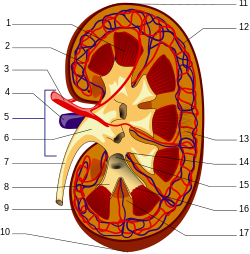
The substance, or parenchyma, of the kidney is divided into two major structures: superficial is the renal cortex and deep is the renal medulla. Grossly, these structures take the shape of 8 to 18 cone-shaped renal lobes, each containing renal cortex surrounding a portion of medulla called a renal pyramid (of Malpighi).[2] Between the renal pyramids are projections of cortex called renal columns (of Bertin). Nephrons, the urine-producing functional structures of the kidney, span the cortex and medulla. The initial filtering portion of a nephron is the renal corpuscle, located in the cortex, which is followed by a renal tubule that passes from the cortex deep into the medullary pyramids. Part of the renal cortex, a medullary ray is a collection of renal tubules that drain into a single collecting duct.
The tip, or papilla, of each pyramid empties urine into a minor calyx, minor calyc


0 comments:
Post a Comment