When looking into someone's eyes, we can easily see several structures:
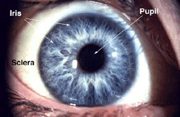
Fig. 1. View of the human eye (59 K jpeg image)
- A colored circular muscle, the iris, which is beautifully pigmented giving us our eye's color (the central aperture of the iris is the pupil). This circular muscle controls the size of the pupil so that more or less light, depending on conditions, is allowed to enter the eye. Eye color, or more correctly, iris color is due to variable amounts of eumelanin (brown/black melanins) and pheomelanin (red/yellow melanins) produced by melanocytes. More of the former is in brown eyed people and of the latter in blue and green-eyed people. The Melanocortin-1 Receptor Gene is a regulator of eumelanin production and is located on chromosome(MCIR) 16q24.3. Point mutations in the MCIR gene will affect melanogenesis. The presence of point mutations in the MCIR gene alleles is a common feature in light skinned and blue/green eyed people (J.A.W. Metzelaar-Blok et al., Invest. Ophthal. Vis. Sci. 42,1951-4, 2001; P. Valverde et al. Nat. Genet. 11, 328-330, 1995)).
- A transparent external surface, the cornea, that covers both the pupil and the iris. This is the first and most powerful lens of the optical system of the eye and allows, together with the crystalline lens the production of a sharp image at the retinal photoreceptor level.
- The "white of the eye", the sclera,which forms part of the supporting wall of the eyeball. The sclera is continuous with the cornea. Furthermore this external covering of the eye is in continuity with the dura of the central nervous system.
When we remove the eye from the orbit, we can see that the eye is a slightly asymmetrical sphere with an approximate sagittal diameter or length of 24 to 25 mm. and a transverse diameter of 24 mm. It has a volume of about 6.5 cc.
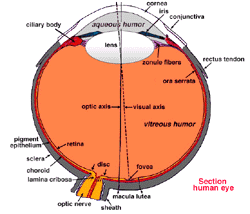
Fig. 2. Sagittal section of the adult human eye(59 K jpeg image)
A cross-sectional view of the eye shows:
- The external layer, formed by the sclera and cornea
- The intermediate layer, divided into two parts: anterior (iris and ciliary body) and posterior (choroid)
- The internal layer, or the sensory part of the eye, the retina
- Three chambers of fluid: Anterior chamber (between cornea and iris), Posterior chamber (between iris, zonule fibers and lens) and the Vitreous chamber (between the lens and the retina). The first two chambers are filled with aqueous humor whereas the vitreous chamber is filled with a more viscous fluid, the vitreous humor.
- The sagittal section of the eye also reveals the lens which is a transparent body located behind the iris. The lens is suspended by ligaments (called zonule fibers) attached to the anterior portion of the ciliary body. The contraction or relaxation of these ligaments as a consequence of ciliary muscle actions, changes the shape of the lens, a process called accommodation that allows us to form a sharp image on the retina.
Light rays are focussed through the transparent cornea and lens upon the retina. The central point for image focus (the visual axis) in the human retina is the fovea. Here a maximally focussed image initiates resolution of the finest detail and direct transmission of that detail to the brain for the higher operations needed for perception. Slightly more nasally than the visual axis is the optic axis projecting closer to the optic nerve head. The optic axis is the longest sagittal distance between the front or vertex of the corna and the furthest posterior part of the eyeball. It is about the optic axis that the eye is rotated by the eye muscles. Some vertebrate retinas have instead of a fovea, another specialization of the central retina, known as an area centralis or a visual streak.


0 comments:
Post a Comment